MMC Sperm (MultiMedia Catalog Sperm) is an automated image analysis software package for sperm quality analysis according to parameters recommended by WHO (World Health Organization). This software is based on our MultiMedia Catalog with addition of dedicated routines for semen analysis. MMC Sperm represents the basis for Computer Assisted Sperm Analysis System (CASA) to be used in human reproduction (in andrology labs, ART, IVF clinics) for estimation of fertility/infertility and understanding its causes as well as in veterinary and animal, bee, fish breeding for calculation of sperm doses for artificial insemination.
Acquisition of images and clips in AVI format from an imaging device provided with DirectShow drivers.
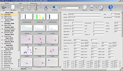
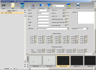
Storage of textual data, images and clips in the built-in MultiMedia Catalog database. You can store patient data, analysis results, video clips, images and any other files in one record (accompanying documents of any office software like MS Office, Open Office etc., Adobe or similar applications for extended image processing). The database provides fast and flexible search and spectacular reporting with images, graphs and other features adjusted to your particular needs. Herewith our database can serve the needs of virtually any laboratory or research institution.
Automated morphology analysis on stained samples according to strict Krueger criteria.
Manual assessment of concentration of white blood cells, immature germ cells, round cells.
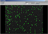
Vitality. Estimation of the percentage of live spermatozoa is assessed by identifying those with an intact cell membrane, from dye exclusion or by hypotonic swelling.
Sperm DNA fragmentation test. Estimation of the percentage of spermatozoa with fragmented DNA (DFI, DNA Fragmentation Index) based on Sperm Chromatin Dispersion method using stains like Halosperm.
Working with camera is much more comfortable than looking into eyepieces (which hurts your eyes by the way).
Using MMC Sperm, you can record all the clips to your hard disk to repeat the motility analysis any time while without it you are restricted to the live time of spermatozoa.
Morphology analysis becomes more accurate. The software will assess every cell that you have acquired.
The use of the automated software for semen analysis helps decreasing the "human factor". It is complicated to analyze, let's say, the form of sperm head by sight while using MMC Sperm you receive objective quantitative data: head length, width, area, form factor. The same applies to motility: estimation of sperm movement becomes more objective if you know its speed and exact track. But the final decision is always yours and you can correct the results of the automated analysis.
Using Computer Aided Sperm Analyzer, it is much easier to train new staff in your laboratory and control the results of their work.
The MultiMedia Catalog database provides the ability to
create unlimited number of records with infinite number of fields to archive all the required patient information accurately and search for it fast.
Having the ability to work over local network, you can select the suitable location of your database (e.g. on the firewall protected server with automatic backup procedures). Even if working on one PC, you are protected from data loss through two-stage deleting procedure in MultiMedia Catalog (if you delete a record, it goes to Recycle Bin first and can be restored).
Selecting proper database field types helps you prevent misprints in your database (you may not enter text into numeric or time fields etc.) Drop-down lists will aid in fast and accurate filling the records with repeatable information. Special tool for insertion of predefined text blocks will aid you in filling the fields with related nomenclature, standard phrases etc.
Your patients will enjoy informative reports with explanations, reference norm values, images (and whatever your creative imagination may produce).
WHY TO SELL
MMC Sperm is the basis for Computer Aided Sperm Analysis System (CASA). Becoming our distributor, you take advantage of being system integrator: sell suitable microscopes, counting chambers, replaceable items, dyes and lots of other complimentary equipment to provide complete customer support. We are always ready to share information where and what to buy at the best prices. We are ready to provide any consultancy on how to integrate a complete CASA and provide extensive customer support including installation and training.
Since we are a team of professional image analysis software developers, we never stand still and continue to improve the software. Software development is never finished and you can count on adding special features required by your customers.
MMC SPERM DATABASE FEATURES
Our database is specially tailored for image analysis needs. You can catalogue virtually any digital assets corresponding with sperm analysis in one place and have fast and smart access to any files.
The volume of semen analysis data, images, clips and documents in your database is restricted only by the size of your storage media.
Your sperm analysis database can be stored on server in your local network which will enable all the security policies and backup procedures provided by Windows. Alternatively, you can also save your spermiogram database to any suitable storage media (internal/external HDD, flash, etc.) if you need to have access to it on different workstations.
Internal archiving tool will help you to keep your important semen analysis and patient data save as well as to make archives with older information to free your storage space.
There are many security tools and procedures to ensure the integrity of your sperm analyzer data. Database records are deleted to internal recycle bin and removed permanently only after you empty your bin. Images and videos are deleted to Windows recycle bin and can be restored. To prevent unwanted changes in the database records, all the records are locked for editing until you press a special button.
To avoid unwanted changes by unauthorized personnel, you can enable PIN mode. The PIN mode will disable the following options until you enter correct PIN: edit database structure (add/delete fields), edit print templates, making changes in the autotext box, deleting database filters, changing default calibrations.
Database structure is implemented as a tree of folders with separate technical files, images, videos, 3rd-party files for every record. This provides the advantage of loading only one record at a time, which makes your work with a large database fast and effective. Furthermore, storage of data in multiple files will protect you in case your PC is infected with a virus: if the virus destroys some files, you will lose only several records and in no case the whole database.
The database is written in XML Unicode which provides wide options of integration of your data in 3rd-party databases as well as the ability to use your local language to fill in the database fields.
Database structure can be easily adjusted by the user through a simple and visually clear interface. Add fields for storing text data (also multiline fields for large amounts of text, drop-down lists with predefined values or automatically generated lists for fast selection of repeating words and phrases), numerical data (e.g. parameter values), use special date tool with calendar. Move single fields or large groups of fields to organize your working space. Rename fields as you need, add any number of fields to store additional sperm analysis data along with automatically calculated parameters.
Use fast search option to find required data in the fields or to retrieve images, video, documents.
Apply extended filters to make selection of records according to any number of parameters (e.g. records created by certain doctor at certain time period having certain diagnosis etc.) Easy filters can be created fast, complicated multiparametric filters can be saved to filter list and reapplied any time.
Numeric fields can be set up to count records in the database automatically according to required parameters (e.g. you can keep track of the number of records with certain diagnosis in your database etc.)
Special autotext box allows you to make an extended tree of predefined text blocks (nomenclature, ready-made phrases describing the diagnosis, prescriptions etc.) Making an advanced tree will let you fill in the patient record with a few clicks without the need of typing long phrases again and again which saves your time and helps to avoid misprints in the record.
Automatic diagnosis. Autotext box also contains special tool which combines database filters and autoinsertion of text blocks to required text fields. Create a filter describing a diagnosis (if a number of parameters meets normal limits) and select this filter as condition of insertion of the diagnosis into corresponding field. This will allow you to provide exact automatic diagnosis with one click without the need to look through a number of parameters and compare them to normal values.
Multilanguage support. If your clinic provides consulting in several languages, using autotext insertion based on filters, you can add diagnosis and other text blocks in as many languages as you need. Add a field marking required language and translate your text with one button.
Database can be displayed in the table mode which allows you to keep track of all the statistics and make your own local reference values of all the measured parameters.
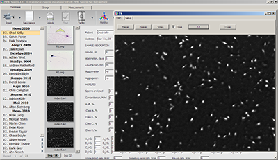
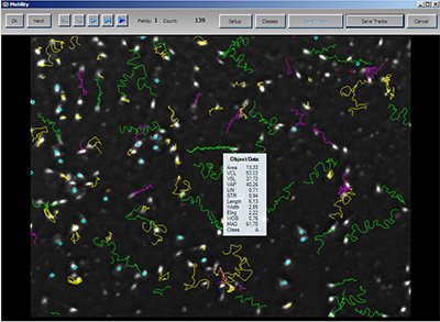
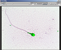
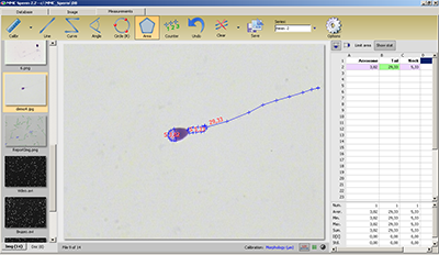
Sperm concentration assessment (M/ml) and motility analysis are performed on native samples by recording AVI clips (acquired live from your imaging device to PC memory or prerecorded to disk). The analysis strictly follows the requirements of the “WHO laboratory manual for the examination of human semen and sperm-cervical mucus interaction”. The analysis is based on frame by frame detection of sperm heads on video clips and building precise tracks which reveal the nature of sperm movement and provide sperm concentration value. The total number of sperms is also calculated automatically. One frame with tracks is automatically saved to database record to serve as visual support in the report or you can save a suitable frame yourself.
The following parameters are calculated:
VCL = curvilinear velocity (micron/s). Time-average velocity of a sperm head along its actual curvilinear path, as perceived in two dimensions in the microscope.
VSL = straight line velocity (micron/s). Time-average velocity of a sperm head along the straight line between its first detected position and its last.
VAP = average path velocity (micron/s). Time-average velocity of a sperm head along its average path. This path is computed by smoothing the actual path.
LIN = linearity. The linearity of a curvilinear path.
STRstraightness. The linearity of the average path.
BCF = beat cross frequency (beats/s). The average rate at which the sperm's curvilinear path crosses its average path.
ALH = amplitude of lateral head displacement. Magnitude of lateral displacement of a sperm head about its average path.
WOB = wobble. A measure of oscillation of the actual path about the average path, VAP/VCL.
MAD mean angular displacement of the sperm head.
Elongation of sperm head.
Mean values of all the parameters are represented in statistics.
Pointing with your mouse cursor to a track, you can see the exact values of the parameters for the selected spermatozoon and classification result in a pop-up window. The clip can be played again, rewound, played frame by frame for detailed analysis. You can save clips to your hard disc uncompressed without loss of quality or by using virtually any codec installed on your PC (xVid codec is recommended). You can adjust detection threshold and also change the classification limits if required.
Based on the parameters mentioned above, the motility of each spermatozoon is graded A, B, C or D (WHO), according to whether it shows:
A = rapid progressive motility
B = slow or sluggish progressive motility
C = nonprogressive motility
D = immotility
A+B = progressive motility
A+B+C = total motility
Additionally, the software enables the user to assess the concentration of:
White blood cells
Immature germ cells
Round cells
Flexible tool for adjustment of sperm head detection will let you work with both bright field and phase contrast (negative phase contrast yields the best results) and not only human sperms but also many animal species.
The number of sperms detected and tracked is displayed on the screen and you can check if you have tracked enough cells (200 according to WHO reference). When the analysis is finished, the results of automated classification and manual counting are automatically transferred to the database.
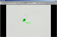
Morphology of the sperm head is an important criterion for the correct diagnosis. The software is set up to analyze still images of smears stained with the Diff-Quik stain according to strict Krueger’s criteria. We have selected Diff-Quik as worldwide recognized leader in rapid staining of sperm. With Diff-Quik, the head is stained pale blue in the acrosomal region and dark blue in the post-acrosomal region which is a good basis for precise image analysis. The following parameters are assessed for every spermatozoon:
Area of the head.
FFC = form factor circle. The degree of similarity of the sperm head to a circle.
Perimeter of the head.
Brightness.
ELL_B = Big axis of ellipse outlining the sperm head, the length of the sperm head.
ELL_S = Small axis of ellipse outlining the sperm head, the width of the sperm head.
Elng = elongation of the sperm head.
FFE = form factor ellipse. The degree of similarity of the sperm head to an ellipse.
Acrosome = Percentage of the acrosomal region.
Mean values of all the parameters are represented in statistics.
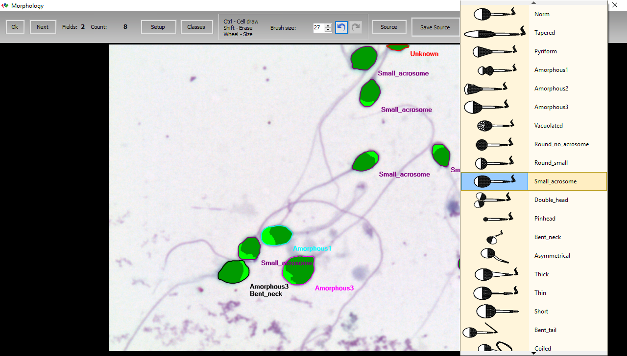
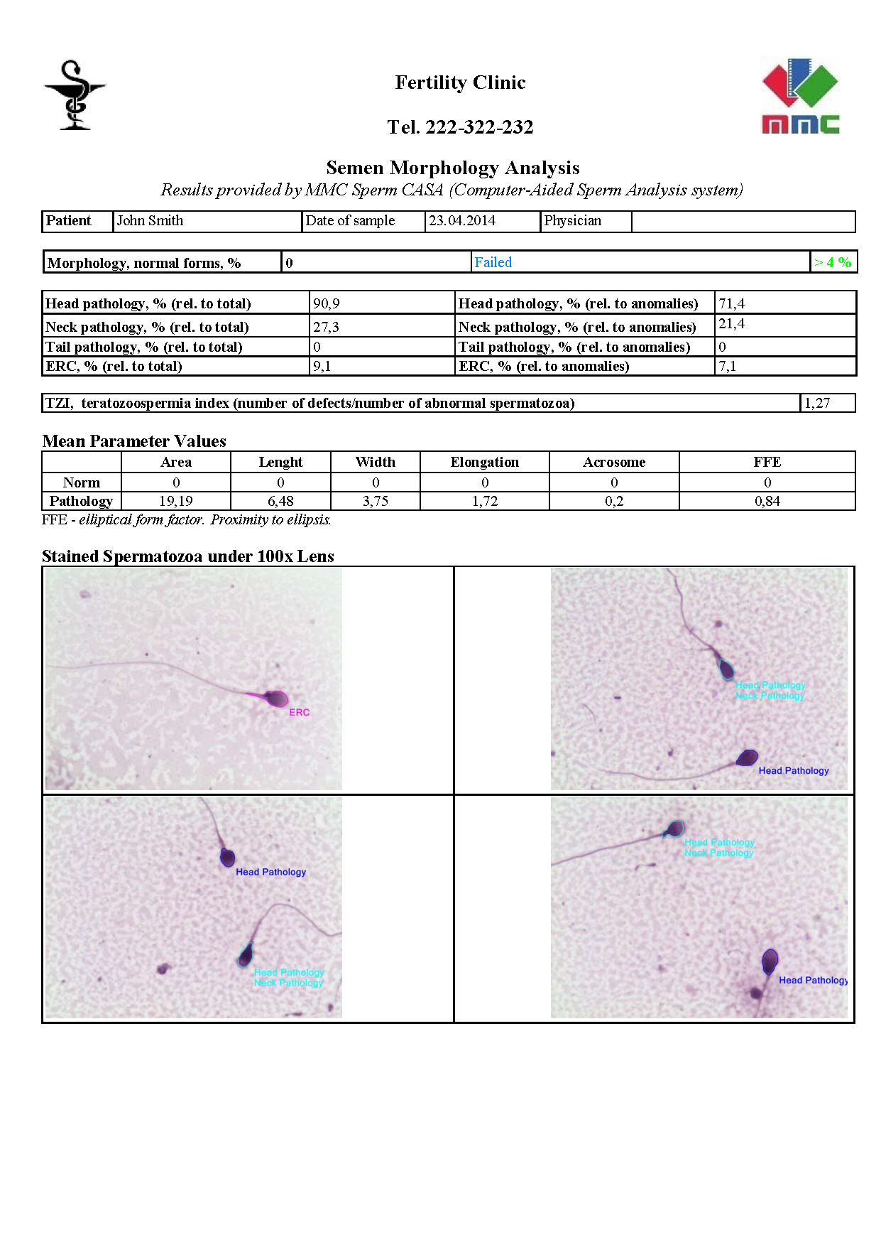
The software classifies spermatozoa into Norm and Head Pathology classes automatically based on head parameters. You can easily correct the results manually and also specify other anomalies (Tail Pathology, Neck Pathology). An extended morphology classifier is available which allows you to specify the following spermatozoa structure abnormalities indicating potential infertility:
tapered, pyriform, round, amorphous, vacuolated, small acrosome, double head, pinhead, bent neck, asymmetrical neck, thick insertion, thin neck, short tail, bent tail, coiled tail, excess residual cytoplasm (ERC).
In case you can not receive a good image of stained smear suitable for automated detection (e.g. because of incorrect sample preparation), there is a special tool which allows you to outline the cells manually.
Software is able to detect both samples prepared according to WHO recommendations for CASA including centrifugation (recommended) but also can be adjusted to easier procedures which provide higher background staining. For complicated cases, there is an option of manual drawing and deleting of objects.
It is now customary to record the number of morphological sperm defects divided by the number of defective spermatozoa, a measure called the teratozoospermia index (TZI). After the analysis is finished, the TZI is calculated automatically. The teratozoospermic index values should read between 1.00 (each abnormal spermatozoon has only one defect) to 3.00 (each abnormal spermatozoon has head, neck and tail defects).
Statistics on all the parameters which have been measured in Motility and Morphology is displayed in the database (mean values of parameters). Statistics provides additional information on sperm quality. Corresponding database fields are filled in automatically during analysis like those for motility and morphology results. Statistics can be printed out to represent it to patient if required. Extended report template should be selected for this purpose (see below). Should the statistical data not be required for the patient, a brief report without statistics can be created.
All the raw data calculated for every spermatozoon can be automatically uploaded to separate text files for your extended research purposes.
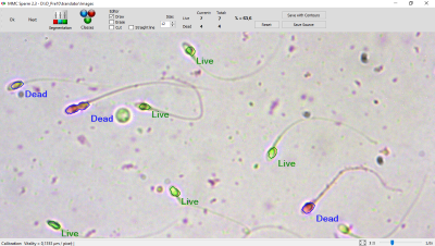
Sperm vitality is an important test, especially for samples with less than about 40% progressively motile sperms. The percentage of live spermatozoa is assessed by identifying those with an intact cell membrane, from dye exclusion or by hypotonic swelling. The dye exclusion method is based on the principle that damaged plasma membranes, such as those found in non-vital (dead) cells, allow entry of membrane-impermeant stains.
The hypo-osmotic swelling test presumes that only cells with intact membranes (live cells) will swell in hypotonic solutions. Sperm vitality should be assessed as soon as possible after liquefaction of the semen sample, preferably at 30 minutes, but in any case, within 1 hour of ejaculation, to prevent observation of deleterious effects of dehydration or of changes in temperature on vitality. It is clinically important to know whether immotile spermatozoa are alive or dead. Vitality results should be assessed in conjunction with motility results from the same semen sample. The presence of a large proportion of vital but immotile cells may be indicative of structural defects in the flagellum. A high percentage of immotile and non-viable cells (necrozoospermia) may indicate epididymal pathology.
Sperm DNA fragmentation test provides additional information on semen fertility potential that can not be achieved by means of standard motility, morphology and vitality tests.
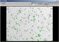
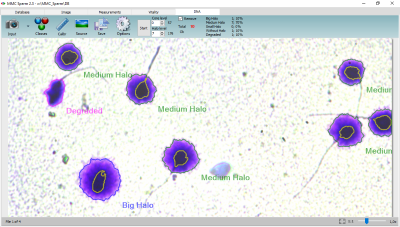
We have selected the Sperm Chromatin Dispersion (SCD) method as simple, fast, accurate, and highly reproducible method for the analysis of sperm DNA fragmentation in semen and processed sperm (Fernández et. al., Journal of Andrology 2003 Jan-Feb;24(1):59-66). The SCD test utilizes the idea that after acid denaturation and removal of nuclear proteins sperm with non-fragmented DNA produce the well seen halo of dispersed DNA loops while spermatozoa with fragmented DNA fail to produce such halo. When somatic cells or spermatozoa with nonfragmented DNA are immersed in an agarose matrix and directly exposed to lysing solutions, the resulting deproteinized nuclei show extended halos of DNA dispersion. The halos correspond to relaxed DNA loops attached to the residual nuclear structure. These deproteinized nuclei are called "nucleoids". The presence of DNA breaks promotes the expansion of the halo of the nucleoid and is the basis for the halo test to detect DNA damage when sperm are treated with an acid solution prior to lysis buffer, the DNA dispersion halos that are observed in sperm nuclei with nonfragmented DNA after the removal of nuclear proteins are either minimally present or not produced at all in sperm nuclei with fragmented DNA. You can use Halosperm staining or contact us for cheaper alternatives.
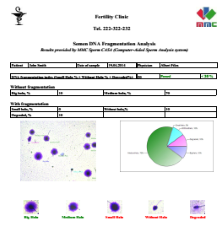
MMC Sperm software allows you to detect and calculate percentage of fragmented sperms automatically. The method is adjusted for the use of bright field 40x lens, no expensive fluorescence staining and equipment required. Spermatozoa are automatically classified into sperms wit Big Halo, Medium Halo, Small Halo, Without Halo and Degraded.
Big Halo
Medium Halo
Small Halo
Without Halo
Degraded
DFI (DNA fragmentation index) is calculated which represents the percentage of fragmented sperms (small halo, without halo, degraded). The DFI below 15% represents fine fertility potential, DFI between 15% and 30% is associated with medium fertility while DFI more than 30% is correlated with bad fertility potential of semen. The distribution of fragmented sperms is represented with bar or pie chart. Special field is used to compare the result to user-defined reference value. For details, see description of measured parameters below.
Manual measurements for special research or other individual tasks are available. You can make measurements on a series of images and send statistical results to previously assigned database fields. Such results will allow you to adjust your classifier limits to any special cases.
Most important image processing operations are available like cropping, image brightness, contrast and color adjustment, rotation and flip, resize, background subtraction etc. Enhance images with filters.
Use any 3rd-party image editors for extended processing and adding comments, figures etc. while still keeping your images in our database. An option allows you to assign external software for editing images and video.
MMC Sperm sperm quality analyzer can be successfully used in veterinary, animal breeding, bee breeding, fish breeding or fish farming as well as in zoos for semen quality control and calcualtion of the number of doses for artificial insemination that can be produced from current sperm sample. Artificial insemination provides several advantages over natural mating: long range of transportation for frozen sperm, more effective use of semen of high quality thoroughbred animals, the use with the purpose of overcoming mating refusal (e.g., in national parks and zoos to breed endangered animal species). Success of artificial insemination directly depends on semen quality, therefore, application of computer assisted sperm analysis in breeding farms is consistently growing all over the world.
MMC Sperm semen analyzer provides special features for calculation of doses. You can set the following parameters in software options:
Dose volume - volume of the semen container to be used (straws, tubes etc.)
Number of sperm per dose - required number of sperms in a dose.
By default, the calculation is based on Motility routine results: percentage of progressively moving sperms (A+B%).
Consider vitality - calculation with respect to percentage of live spermatozoa in the sample if sperm vitality has been tested.
Consider morphology - calculation with respect to percentage of sperms with normal morphology if sperm morphology has been tested.
By the end of the semen quality analysis, the software calculates corresponding number of doses for artificial insemination and volume of extender that should be added to current sperm sample to get this number of doses.
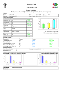
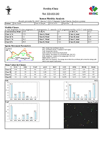
All the data you have provided in the patient record along with automatically calculated parameters, corresponding bar or pie charts and images can be placed into your report (spermiogram). Spermiograms are based on *.rtf format which is recognized by virtually any text editors. You can integrate your spermiogram into any local document form or use our default templates. Internal document editor will allow you to create your own *.rtf files, save the resulting reports to corresponding records. Create as many templates as you need for your clinic: brief ones containing only text and prescriptions, full reports with all the statistical data and images, comprehensive charts and visually supported explanations to patient’s diagnosis. If you have some special requirements, please contact us. We will help to adjust the report.
Since MMC Sperm acts as a part of Computer Aided Sperm Analysis system, there are some special hardware requirements that you should consider. Generally, it is not so complicated to gather all the components of the system by yourself but we highly recommend contacting us to ask for a local distributor in your area to get all the equipment from one hand. If there is no distributor in your area, we will be happy to share direct contacts to reliable hardware manufacturers that we have collected during the years of our work. Our target is to earn on our software product, therefore we are ready to provide you any help in buying other hardware parts at the best price. Details on microscope, camera, PC selection for MMC Sperm.
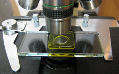
A Computer Aided Sperm Analyzer (CASA) consists of a PC with sperm analysis software installed, a microscope with imaging device attached to it and a special sperm counting chamber.
PC requirements are mostly determined by the need of recording high resolution video clips at 60 fps speed in motility analysis. Other analyses are not that demanding. If you already have a modern PC that you would like to use, the first step would be ordering the required digital camera. MMC Sperm software provides a tool to test real PC performance with camera even in demo software mode. If you want to buy a new PC, follow the instructions below:
64 bit Windows 10 (Windows 7, 8 supported but no recommended).
If possible, avoid laptops since their components provide less performance, use desktop PCs.
Intel chipsets (no AMD chipsets), chipsets with Z mark recommended, Z270 and higher recommended. Check currently available processors and chipsets on Intel web page
Intel i5, i7, i9, Xeon processors, 7th generation and higher, Intel HD Graphics 630 and higher.
Minimum 8 Gb operating memory, DDR4 recommended.
If you have to buy a laptop, look among "gamers" laptops, they provide better components. Look for Intel processors of 7th generation and higher with HQ or HK mark. Make sure the graphics of your processor is Intel HD Graphics 630 and higher. Always check real specifications of selected laptop model on manufacturer's web site. Recommended mobile processors:
Special counting chambers with fixed depth should be used for motility analysis: Leja, Makler, MicroCell, Cell-Vu etc. Such chambers provide standard volume and monolayered distribution of sperms in one focal plane. We offer a handy and cost-effective alternative to well-known Makler counting chamber: reusable MMC-SK sperm counting chamber.
Leja counting slides are useful for a Computer Aided Sperm Analysis System if the clinic or laboratory has got a large volume of analyses.
Leja offers a wide range in disposable counting chambers for standard semen analysis. There are slides with 2 or 4 chambers and with chamber depths of 10 or 20 micron. The Leja slides have a standard low level of quantification which implies that even low sperm counts can be determined. The clinical implication of this is that you are well equipped to determine a threshold to choose between IUI, IVF, or ICSI for example. Leja slides are very easy to work with and allow a laboratory technician to quickly learn to perform an accurate count thus improving efficiency and quality.
Trinocular upright transmitted light laboratory grade microscope to mount camera on it. Beam splitter 100/0 (all light should be transferred to camera).
Camera adapter corresponding with image sensor size. We recommend 1/2"-1/1.8" sensor with 1.0x adapter. Larger sensors might be used while applying Region of Interest in camera drivers in motility mode since with 10x objective the sperms might appear too small for comfortable quality control.
You can choose bright field or phase contrast. MMC Sperm software is able to detect both dark objects on light background and vice versa. Negative phase contrast provides the best results! Our MMC-SK counting chamber due to its transparent glass base unit works very well with phase contrast even on condensers with standard working distance.
10x lens for motility. 10x objective provides required focus depth. 20x lenses usually can not get all sperms in focus even with 10 micron deep chambers which disturbs sperm detection. If you want to use additional 20x lens for manual sperm count on counting chambers like the one from Makler, a lens with long working distance should be used. In case of disposable chambers like Leja, there is no need in long WD 20x lens. We recommend using negative phase contrast: sperm heads are covered with light accurately, the background is dark, a lot of debris is being eliminated. Positive phase contrast and bright field are available but have their downsides. Positive phase contrast does not cover the whole sperm head which does not allow us to use head form parameters to differentiate sperms from debris. Furthermore, positive phase contrast might provide halos around sperm heads which can be detected as additional sperms by the software. Bright field makes it harder to differentiate between sperms and debris.
100x oil bright field lens for morphology.
40x bright field lens for vitality and DNA fragmentation analysis.
Light source: 30W halogen (or higher). Equivalent LED is highly recommended since halogen lamps might provide flickering at 60 fps if microscope power unit is not stabilized well enough.
Stage micrometer - calibration scale or graticule. A transparent slide with 1mm scale with 100 divisions 0.01mm.
We are continously looking for the best hardware solutions for sperm analysis systems, develop direct drivers. Please contact us for currently recommended digital camera. There are several reasons why only recommended digital cameras should be used with MMC Sperm software:
We test cameras for suitability, performance and reliability. Cameras that we recommend keep fine balance between image sensor quality, speed and cost-effectiveness.
For sperm motility, image clips with 1.3 M resolution and higher at minimum of 60 fps are analyzed directly from PC memory without recording to disk. This provides the maximum performance on minimum of PC hardware. Using third party cameras, you will have to record clips to disk which decreases the analysis speed.
The camera has got the ability to record all the settings you have applied for motility, morphology, vitality and DNA fragmentation as well as load them automatically when corresponding analysis starts.
Our direct drivers provide flexible set up of all essential camera parameters which allows you to solve both standard routine and complicated research tasks.
For details on sample preparation protocol, please refer to the
WHO laboratory manual for the examination and processing of human semen (5th edition 2010). This manual provides updated, standardized, evidence-based procedures and recommendations for laboratory managers, scientists and technicians to follow in examining human semen in a clinical or research setting. Detailed protocols for routine, optional and research tests are elaborated. Visit WHO web page for printed version.
Motility
Sperm concentration is estimated using special counting chambers with fixed depth to provide standard volume and distribute spermatozoa in one focal plane to let them move free and stay in focus all the time they are being tracked. Sperm counting chambers provide the ability to work with undiluted specimen. The preparation consists usually in mixing the sperm sample carefully while avoiding bubbles. If the specimen is highly mucous, it can be aspirated with syringe several times to make it more uniform.
To achieve good quality images of stained spermatozoa for morphology assessment, seminal plasma should be diluted and removed after centrifugation. The sperm pellet is resuspended in an
appropriate volume to obtain the highest sperm concentration possible, but not exceeding 80 x 10^6/ml. An aliquot of 0.2 to 0.5 ml of semen, depending on sperm concentration, is diluted to 10 ml with normal saline at room temperature. The tube is centrifuged at 800g for 10 minutes and most of the supernatant tipped off. The pellet is resuspended in the remaining saline, typically 20-40 µl, by gently tapping the tube. Then 5-10 µl of this suspension is placed on a glass microscope slide and the drop is spread across the slide with a pipette to make a smear as described below.
Making smears for staining for sperm morphology. (a) Feathering method for undiluted
semen. The semen drop (S) spreads along the back edge of the angled slide and is pulled forward over the flat slide to form the smear. (b) Pipette method for washed samples. A drop of the suspension (S) is spread over the surface of the slide by the horizontally held pipette (P).
Preparation of smears. At least two smears should be made from the fresh semen sample for duplicate assessment and in case of problems with staining. The slides should first be thoroughly cleaned, washed in 70% ethanol and dried, before a small drop of semen (5 to 20 µl) is applied to the slide. If the sperm concentration is over 20 x 10^6/ml, then 5 µl of semen can be used: if the sperm concentration is less than 20 X 10^6/ml, then 10 to 20 µl of semen should be used. The 'feathering' technique whereby the edge of a second slide is used to drag a drop of semen along the surface of the cleaned slide may be used to make smears of spermatozoa, but care must be taken not to make the smears too thick. Feathering works well when viscosity is low but is often unsuitable for viscous semen. Alternatively, a drop of semen can be placed in the middle of a slide and then a second slide, face down, placed on top so that the semen spreads between them: the two slides are then gently pulled apart to make two smears simultaneously.
The software is adjusted to be used with Diff-Quik staining. Diff-Quik is a rapid staining kit recommended by WHO and popular worldwide. Rapid staining methods are particularly useful for clinical laboratories that have high throughput. A Diff-Quik kit consists of:
Fixative reagent (triarylmethane dye dissolved in methanol).
Staining solution 1, red (eosinophilic xanthene).
Staining solution 2, blue (basophilic thiazine).
How to use Diff-Quik:
Immerse slides prepared as described above in triarylmethane fixative for 15 seconds.
Drain the excess solution by placing slides vertically on absorbent paper.
Immerse in rapid stain solution 1 for 10 seconds.
Drain the excess solution by placing slides vertically on absorbent paper.
Immerse in rapid stain solution 2 for 5 seconds.
Immerse the slide in running tap water 10 to 15 times to remove excess stain.
To achieve a good staining, please follow the instructions supplied with your Diff-Quik kit!
Vitality
MMC Sperm semen quality analyzer provides two modes for sperm vitality analysis: automated and manual. In manual mode any available staining can be used: eosin, eosin-nigrosin, hypo-osmotic swelling (HOS) or fluorescence staining. Automated sperm vitality analysis method is based on eosin alone (Eosin Y, colour index 45380). This is the fastest and cheapest method of spermatozoa staining. Preparation of reagents:
NaCl, 0,9% (w/v): dissolve 0,9 g NaCl in 100 ml purified water.
Eosin Y, 0.5% (w/v): dissolve 0.5 g of eosin Y (colour index 45380) in 100 ml of 0.9% NaCl.
Some commercially available eosin solutions are hypotonic aqueous solutions that will stress the spermatozoa and can yield false-positive results. To use such a solution, add 0.9 g of NaCl to 100 ml of solution to raise the osmolality.
How to prepare vitality staining:
Mix the sample well but avoid bubbles.
Mix 5 microliter of semen with 5 microliter of eosin solution.
Aspermia - no semen (no or retrograde ejaculation).
Asthenozoospermia - percentage of progressively motile spermatozoa (A+B classes) below the lower reference limit.
Asthenoteratozoospermia - percentages of both progressively motile (A+B classes) and morphologically normal spermatozoa below the lower reference limits.
Azoospermia - no spermatozoa in the ejaculate.
Cryptozoospermia - spermatozoa absent from fresh preparations but observed in a centrifuged pellet.
Haemospermia (haematospermia) - presence of erythrocytes in the ejaculate.
Leukospermia (leukocytospermia, pyospermia) - presence of leukocytes in the ejaculate above the threshold value.
Necrozoospermia - low percentage of live, and high percentage of immotile, spermatozoa in the ejaculate.
Normozoospermia - total number or concentration of spermatozoa, and percentages of progressively motile (A+B classes) and morphologically normal spermatozoa, equal to or above the lower reference limits.
Oligoasthenozoospermia - total number or concentration of spermatozoa, and percentages of progressively motile (A+B classes), below the lower reference limits.
Oligoasthenoteratozoospermia - total number or concentration of spermatozoa, and percentages of both progressively motile (A+B classes)and morphologically normal spermatozoa, below the lower reference limits.
Oligoteratozoospermia - total number or concentration of spermatozoa, and percentage of morphologically normal spermatozoa, below the lower reference limits.
Oligozoospermia - total number or concentration of spermatozoa below the lower reference limit.
Teratozoospermia - percentage of morphologically normal spermatozoa below the lower reference limit.
Our software measures a large set of semen quality parameters which we update on the regular basis. To send a parameter value to your database you have to create a numeric field with certain reserved field ID (internal field name that is being given while you create a new field, not to be confused with field label, which you can edit in field properties anytime). So, to add a new automeasurement result, you not only need to update the software (during updates, your database is not being changed automatically for security purposes) but also to add a new numeric field with given field name. This field starts to receive new measurement results. The table below represents the list of actual parameters being measured by the software and corresponding database fields.
Parameter
Field ID
Volume - sample volume. This value is used to calculate the overall number of sperms in the sample. Should be filled in by the user before analysis.
Volume
Motility
Sperms analyzed - the number of sperms that have been analyzed during the work of the Motility routine. Is used for quality control - according to WHO requirements 200-400 sperms should be analyzed.
CountMotility
Fields analyzed the number of microscope fields analyzed duringMotility routine.
Used for quality control. At least 4 fields in different spots of the counting chamber recommended (however, priority should be given to the minimum number of analyzed sperms, see above).
MotilityFrames
Concentration M/ml - sperm concentration in millions per milliliter.
Concentration
Status concentration - the result of comparison to normal reference values given in the software options. Field values - Passed or Failed.
statusConc
Total number of sperms in the current sample. Is calculated based on the value of the Volume field. Should the Volume field be empty, the total number is not being calculated. Make sure to fill in sample volume before analysis!
TotalCount
Status total number - the result of comparison to normal reference values given in the software options. Field values - Passed or Failed.
statusTotal
A+B, % - percentage of progressively motile sperms.
A_plus_B
Status А+В - the result of comparison to normal reference values given in the software options. Field values - Passed or Failed.
statusPR
A+B+C, % - percentage of motile sperms in the sample including nonprogressively motile ones.
ABC
Status А+В+С - the result of comparison to normal reference values given in the software options. Field values - Passed or Failed.
statusPR_NP
Class A, % - percentage of sperms moving fast and straight.
Class_A
Class A, M/ml - concentration of sperms moving fast and straight.
A_conc
Class A, total - number of sperms moving fast and straight.
A_total
Class B, % - percentage of sperms moving slow but straight.
Class_B
Class B, M/ml - concentration of sperms moving slow but straight.
B_conc
Class B, total - number of sperms moving slow but straight.
B_total
Class C, % - percentage of nonprogressively moving sperms.
Class_C
Class C, M/ml - concentration of nonprogressively moving sperms.
C_conc
Class C, total - number of nonprogressively moving sperms.
C_total
Class D, % - percentage of immotile sperms.
Class_D
Class D, M/ml - concentration of immotile sperms.
D_conc
Class D, total - number of immotile sperms.
D_total
A_VCL - average value of VCL parameter among spermatozoa of class A .
A_VCL
A_VSL - average value of VSL parameter among spermatozoa of class A.
A_VSL
A_VAP - average value of VAP parameter among spermatozoa of class A.
A_VAP
A_ALH - average value of ALH parameter among spermatozoa of class A.
A_ALH
A_BCF - average value of BCF parameter among spermatozoa of class A.
A_BCF
A_LIN - average value of LIN parameter among spermatozoa of class A.
A_LIN
A_STR - average value of STR parameter among spermatozoa of class A.
A_STR
A_WOB - average value of WOB parameter among spermatozoa of class A.
A_WOB
A_MAD - average value of MAD parameter among spermatozoa of class A.
A_MAD
B_VCL - average value of VCL parameter among spermatozoa of class B.
B_VCL
B_VSL - average value of VSL parameter among spermatozoa of class B.
B_VSL
B_VAP - average value of VAP parameter among spermatozoa of class B.
B_VAP
B_ALH - average value of ALH parameter among spermatozoa of class B.
B_ALH
B_BCF - average value of BCF parameter among spermatozoa of class B.
B_BCF
B_LIN - average value of LIN parameter among spermatozoa of class B.
A_LIN
B_STR - average value of STR parameter among spermatozoa of class B.
B_STR
B_WOB - average value of WOB parameter among spermatozoa of class B.
B_WOB
B_MAD - average value of MAD parameter among spermatozoa of class B.
B_MAD
C_VCL - average value of VCL parameter among spermatozoa of class C.
C_VCL
C_VSL - average value of VSL parameter among spermatozoa of class C.
C_VSL
C_VAP - average value of VAP parameter among spermatozoa of class C.
C_VAP
C_ALH - average value of ALH parameter among spermatozoa of class C.
C_ALH
C_BCF - average value of BCF parameter among spermatozoa of class C.
C_BCF
C_LIN - average value of LIN parameter among spermatozoa of class C.
C_LIN
C_STR - average value of STR parameter among spermatozoa of class C.
C_STR
C_WOB - average value of WOB parameter among spermatozoa of class C.
C_WOB
C_MAD - average value of MAD parameter among spermatozoa of class C.
C_MAD
D_VCL - average value of VCL parameter among spermatozoa of class D.
D_VCL
D_VSL - average value of VSL parameter among spermatozoa of class D.
D_VSL
D_VAP - average value of VAP parameter among spermatozoa of class D.
D_VAP
D_ALH - average value of ALH parameter among spermatozoa of class D.
C_ALH
D_BCF - average value of BCF parameter among spermatozoa of class D.
D_BCF
D_LIN - average value of LIN parameter among spermatozoa of class D.
D_LIN
D_STR - average value of STR parameter among spermatozoa of class D.
D_STR
D_WOB - average value of WOB parameter among spermatozoa of class D.
C_WOB
D_MAD - average value of MAD parameter among spermatozoa of class C.
D_MAD
White blood cells, M/ml - concentration of white blood cells calculated manually in Motility routine.
Vitality, sperms analyzed - number of sperms analyzed during the work of theVitality routine. Is used for quality control. Minimum of 200 spermatozoa should be analyzed.
VitTotal
Vitality, % - % of live sperms. Result of the Vitality routine.
Vitality
Dead, % - % of dead sperms. Result of the Vitality routine.
Dead
Status vitality the result of comparison to normal reference values given in the software options. Field values - Passed or Failed.
statusVit
Total live number the number of live spermatozoa in the specimen. Calculated based on the number in the Total number field, Motility section.
live_total
Morphology
Sperms analyzed - number of sperms that have been processed during the work of the Morphology routine. Is used for quality control - according to WHO requirements 200-400 sperms should be analyzed.
CountMorphology
Norm, % - percentage of spermatozoa with normal morphology as per assessment in Morphology routin.
Norm
Status morphology - the result of comparison to normal reference values specified in software options. Field values - Passed or Failed.
statusMorph
In the Morphology routine, all pathological forms are calculated in two versions % rel. to total (percentage of spermatozoa with given pathology relative to total number of sperms that have been analyzed) and % rel. to anomalies (percentage of spermatozoa with given pathology relative to all pathological sperms that have been analyzed, is used to highlight prevailing pathology in given sample). Field IDs of % rel. to anomalies differ by added P letter.
% rel. to total parameters, simplified classifier:
Head pathology.
HeadPathology
Neck pathology.
NeckPathology
Tail pathology.
TailPathology
ERC - excess residual cytoplasm.
ERC
% rel. to anomalies, simplified classifier:
Head pathology.
HeadPathologyP
Neck pathology.
NeckPathologyP
Tail pathology.
TailPathologyP
ERC - excess residual cytoplasm.
ERCP
% rel. to total parameters, extended classifier:
Tapered heads
Tapered
Pyriform heads
Pyriform
Amorphous heads, type 1 - amorphous pyriform.
Amorphous1
Amorphous heads, type 2 - amorphous tapered.
Amorphous2
Amorphous heads, type 3 - amorphous oval.
Amorphous3
Vacuolated heads
Vacuolated
Round heads, no acrosome
Round_no_acrosome
Round small heads
Round_small
Heads with small acrosome
Small_acrosome
Double heads
Double_head
Pinheads
Pinhead
Bent necks
Bent_neck
Necks with asymmetrical insertion
Asymmetrical
Thick necks
Thick
Thin necks
Thin
Short tails
Short
Bent tails
Bent_tail
Coiled tails
Coiled
% rel. to anomalies parameters, extended classifier:
Tapered heads
TaperedP
Pyriform heads
PyriformP
Amorphous heads, type 1 - amorphous pyriform.
Amorphous1P
Amorphous heads, type 2 - amorphous tapered.
Amorphous2P
Amorphous heads, type 3 - amorphous oval.
Amorphous3P
Vacuolated heads
VacuolatedP
Round heads, no acrosome
Round_no_acrosomeP
Round small heads
Round_smallP
Heads with small acrosome
Small_acrosomeP
Double heads
Double_headP
Pinheads
PinheadP
Bent necks
Bent_neckP
Necks with asymmetrical insertion
AsymmetricalP
Thick necks
ThickP
Thin necks
ThinP
Short tails
ShortP
Bent tails
Bent_tailP
Coiled tails
CoiledP
TZI/MAI along with simplified classifier - TZI (1-4 anomalies per spermatozoon), along with extended classifier - MAI (can be more than 4 anomalies).
TZI
Area - average value of Head area parameter for normal spermatozoa.
N_Area
Length - average value of Head length parameter for normal spermatozoa.
N_Lenght
Width - average value of Head width parameter for normal spermatozoa.
N_Widht
Elongation - average value of Head elongation parameter for normal spermatozoa.
N_Elongation
Acrosome - average value of Acrosome % parameter for normal spermatozoa.
N_Acrosome
Form Factor Ellipse - average value of FFE parameter for normal spermatozoa.
N_FFE
Area - average value of Head area parameter for pathological spermatozoa.
P_Area
Length - average value of Head length parameter for pathological spermatozoa.
P_Lenght
Width - average value of Head width parameter for pathological spermatozoa.
P_Widht
Elongation - average value of Head elongation parameter for pathological spermatozoa.
P_Elongation
Acrosome - average value of Acrosome % parameter for pathological spermatozoa.
P_Acrosome
Form Factor Ellipse - average value of FFE parameter for pathological spermatozoa.
P_FFE
DNA Fragmentation
DNA, sperms analyzed - number of sperms analyzed during the work of the DNA Fragmentation routine. Is used for quality control. Minimum of 500 spermatozoa should be analyzed.
DNA_total
Big halo, % - percentage of spermatozoa producing big halo after treatment by Sperm Chromatin Dispersion method during the work of the DNA Fragmentation routine. Sperms with big halo are considered nonfragmented.
BigHalo
Medium halo, % - percentage of spermatozoa producing medium halo after treatment by Sperm Chromatin Dispersion method during the work of the DNA Fragmentation routine. Sperms with medium halo are considered nonfragmented.
MediumHalo
Small halo, % - percentage of spermatozoa producing small halo after treatment by Sperm Chromatin Dispersion method during the work of the DNA Fragmentation routine. Sperms with small halo are considered those with fragmented DNA.
SmallHalo
Without halo, % - percentage of spermatozoa not producing any halo after treatment by Sperm Chromatin Dispersion method during the work of the DNA Fragmentation routine. Sperms without halo are considered those with fragmented DNA.
WithoutHalo
Degraded, % - percentage of spermatozoa not producing any halo and having pale staining after treatment by Sperm Chromatin Dispersion method during the work of the DNA Fragmentation routine. Sperms without halo are considered those with fragmented DNA.
Degraded
DFI - DNA Fragmentation Index, percentage of spermatozoa with fragmented DNA: smal halo + without halo + degraded. The result of the work of the DNA Fragmentation routine.
DFI
Status DNA the result of comparison to normal reference values given in the software options. Field values - Passed or Failed.
Doses - number of doses which can be prepared from current sample. Dose volume and required number of sperms per dose can be specified in software option.
doses
Volume of diluent - volume of extender (diluent) that should be added to get the required number of doses calculated in Doses field.