|
|||||||
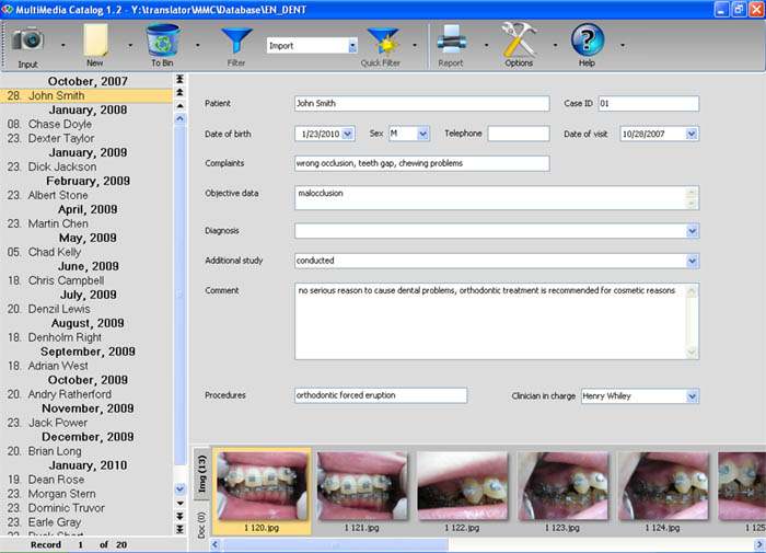
Intraoral and extraoral digital imaging to control teeth treatment has already become everyday practice in dentist office. Patients associate a modern dental clinic with high technology, cutting edge medical equipment, high level of comfort. And a photo archive of images taken during the treatment would be an attractive and useful addition to the dentist's equipment. Intraoral imaging not only aids in controlling the clinical situation and makes the analysis easier, but also improves the patient's motivation and trust to his dentist. It is not a big secret that visual representation of any information is the most effective one. This is why people use visualization as the most convincing way to create an image of desired reality. If you want your idea to be perceived easily and completely, visualize it! Dentists meet this challenge in their everyday practice. If the teeth treatment is caused by pain, or serious objective reasons, there is no additional motivation required, but any procedure which is made for cosmetic reasons require extra motivation of your patient which e.g. is often the case in orthodontics. And digital photographs are the most impressive way to decision making. With some minor exceptions, a stomatologist is not a professional in digital photography and image processing, therefore such issues as selecting the right digital camera, acquiring images and subsequent image processing, archiving and retrieving are quite actual. The solution of the problems which begin after the picture has been taken is not as easy to find as it looks. While image processing issues can be solved by means of such popular software packages as Adobe Photoshop, Corel Draw, Photo Editor etc., image archiving for stomatological purposes is a special topic. An intraoral image is just a part of a collection of data which is required for patient treatment: results of radiodiagnosis, tomography, treatment plan etc. All of these files are usually stored separated from photo archive which makes the retrieval and analysis of all the required patient information complicated and time-consuming. MMCSoft offers a solution to all of these systematization and interconnection problems MultiMedia Catalog Dent (MMC Dent).
|
|||||||
|
|||||||
 |
|||||||
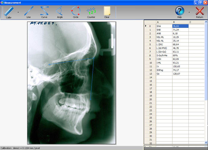
The main purpose of MMC Dent is convenient file management to reduce the unnecesary work load of the dentist connected with image processing and archiving issues and herewith to make the stomatological practice more efficient and comfortable for both the stomatologist and the patient. Special measurement feature allows the doctor to analyze any radiographic images (lateral and frontal cranial x-rays, panoramic dental x-rays) by using calibrated angular and linear measurements in real units. Stomatologist can customize the output by combining different parameters. Furthermore, there are two dedicated modes for lateral cephalometric analysis of radiographs and soft tissue analysis. The user places landmarks on the image, the software calculates required parameters immediately. |
|||||||
| Parameters in cephalometry mode | |||||||
| SNA, SNB, ANB, SN-SNАPNS, AiAr-NS, BiBr-OrPo, AiBiMp-ML, 1-SN, 1-ML, AiAr-BiBr, Go, NSL-NL, NSL-ML, Y-axis, NL-ML, SN-GoGn, ANSPNS-GoGn, AiAr-ANSPNS, AiAr-SN, BiBr-GoGn, wits, SN, A-ANSPNS, ANS-PNS, Go-Gn, R1-R2, R1R2/GnGo, NSAr, SArGo, ArGoMe, SGo/NMe, BaN-SMe. | |||||||
| Parameters in soft tissue profile mode: | |||||||
|
|||||||
| MMC Dent also displays reference Norm values of the calculated parameters. Results of cephalometric analysis as well as soft tissue profile analysis are automatically saved to database. Parameter values can also be exported to third party software for further statistical treatment via Windows Clipboard. DOcuments of third-party software (e.g. Excel sheets) can be created and stored directly in the corresponding database record! | |||||||
 |
|||||||
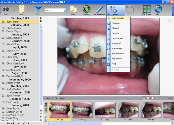
If the included set of image processing tools like auto contrast, sharpening, smoothing etc. is not enough, for sophisticated processing the dentist can easily export image to favorite external software and then update changes in the database. |
|||||||
 |
|||||||
| Back to top | |||||||
| Request demo version | |||||||

| © 2019 MMCSoft. All rights reserved. | ||||||