 |
 |
 |
|
MMC MultiMeter software for microscopy and machine vision |
|
| |
|
|
| |
MMC MultiMeter is a universal microscope software, software for machine vision and quality control: automatic object detection, classification and statistical treatment of series of images. |
|
| |
MMC MultiMeter comprises all the functions of the MultiMedia Catalog database with additional set of tools for image analysis: object detection and classification, statistical analysis of series of images with bar charts and dependency graphs. MMC MultiMeter can be applied in medicine (histology, cytology), life science, material science (particle analysis, phase analysis), food industry, production quality control etc. We can provide complete solution and recommend you a camera from our product line of digital microscope cameras or produce a customized solution for you. Contact us and provide detailed information on your application, we will offer tailored software and hardware solution to your task. |
|
| |
|
|
| |
|
|
| |
- Image segmentation by brightness and color.
- Convenient editor for object correction, separation and manual drawing.
- Operations with object masks to complete detection: opening, closing mask contours, dilation and erosion.
- Automated measurement of a number of parameters like form, dimensions, optical features.
- Data display on every measured object.
- Classification: automatic by a parameter or by manually defined classes.
- Classifier training by objects.
- Statistical treatment and accumulation of measurement results for series of images.
- Adjustable bar charts and data output on charts.
- Sending measurement results to database.
- Saving bar charts as images to database.
- Sending measurement results to Excel, Word and other application.
- Print out according to customizable templates.
- Simple visual set up of analysis algorithms for easy automation of image analysis.
- We offer MMC MultiMeter with our digital microscope cameras, which are controlled by embedded direct drivers.
|
|
|
|
| |
|
|
| |
 Object Detection (Segmentation, Binarization) Object Detection (Segmentation, Binarization)
|
|
| |
|
|
| |
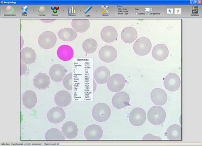
In our MMC MultiMeter microscope software, we have implemented two tools for object detection: a simple one where you point to the image areas that belong to the objects of your microscopic analysis and an enhanced tool for image segmentation based on both color and greyscale levels.
|
|
| |
|
|
| |
 |
|
 |
|
 |
| Source image |
|
Image with object masks |
|
Results of statistical treatment |
|
|
| |
|
|
| |
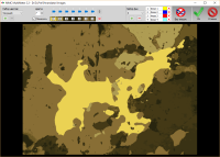
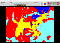
Object detection tool is crucially important for automated image analysis. If the automatic objects detection turns out to be not possible, and too much manual correction is required, this could mean, the image analysis task is not efficiently solved or not solvable at all. Therefore we have done our best to make our object detection as flexible and intelligent as possible. Our extended tool for image segmentation detects the given number of colors or greyscale levels on the image you acquired from microscope. To control the number of required color/greyscale levels you can enable a mode that shows you the segmented image "with the eyes of the software". It allows you to determine whether the selected number of levels is necessary and sufficient for current objects: in simple cases, you would only need 2-3 levels to be detected, in case of complicated multicolored objects you need more levels. You should tick the colors or grey levels that correspond with the objects of your microscopic investigation or click on objects in the image. This covers the selected image areas that should be considered as objects with masks. You can switch between different colors of the masks to get contrasted mask view with regard to image colors. Fine tuning of the weight of every detected component is available using sliders or mouse wheel. Background correction mode can be used to eliminate noisy background. Color or greyscale level sets can be saved for future use on similar microscope specimens.
|
|
| |
|
|
| |
 |
| |
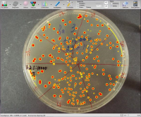
| Assessment of bacterial colonies in Petri dish: the dish has been outlined with a frame while the area outside the frame is not being analyzed. |
|
|
| Some tasks require restriction of the area in which the analysis is being performed. E.g., estimation of growth of bacteria colonies respective to Petri dish in which they are placed. MMC MultiMeter provides a frame drawing tool to define the image segmentation area. Furthermore, complex areas selected with object detection tool can be used as multiple frame. |
| |
Phase analysis
If objects of microscopic analysis are not particles of various shapes but extended areas of the image with large area and complex shapes and types of objects are known in advance (e.g., rock structure components in geology, stained tissue areas in histology), MMC MultiMeter offers special mode uniting detection and classification - phase analysis. The user provides the number and names of classes and assigns detected image areas to object classes. There is also a tool for applications like NCR (nuclear-cell ratio, object inside another object). |
|
|
|
| |
|
|
| |
 Processing Object Masks Processing Object Masks
|
|
| |
|
|
| |
|
|
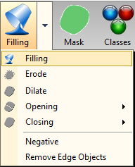
Despite the presence of a flexible tool for object detection, sometimes additional processing of object masks is required to cover the areas of investigation with most precision. The following operations with object masks are implemented in MMC MultiMeter:
-
Filling the areas inside the object boundaries. For cases when some areas inside objects have characteristics close to surrounding background and their detection leads to simultaneous selection of large background areas.
-
Erosion removes one layer of pixels on object perimeter. Can be used to remove small noise and uneven object boundaries or to restore object boundaries after application of Dilation.
-
Dilation adds one layer of pixels on object perimeter. Is effective in closing small spaces between objects or between separated parts of one object. Can also be used to restore objects after application of Erosion.
-
Opening and Closing are two complex operations combining erosion and dilation. Opening involves the given number of erosions followed by the same number of dilations. Closing, vice versa, involves the given number of dilations followed by the same number of erosions. These operations result in cleaner object boundaries without noise (smaller masks around object perimeter).
-
Negative - invert object masks, i.e., areas that were considered as objects become background.
- Remove Edge Objects - removes all the objects that touch the edge of the image. Form and dimensions of such objects are unknown since parts of them are missing on the image. Such objects usually should not be considered in measurements and statistics.
|
|
|
| |
|
|
| |
 Object Classification Object Classification
|
|
| |
|
|
| |
|
|
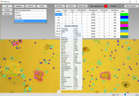
After detection of objects on microscope image and measurements of their parameters, next step is classification of objects. Two types of classifiers are implemented in MMC MultiMeter: classification based on known class limits and statistical classifier. When objects of microscopic analysis are classified by known limits, the user chooses required parameters and inputs the values dividing the classes. This type of classifier can be trained by assigning classes to objects on image. If a statistical classifier is used, the software utilizes mean values of the selected parameters and their dispersion. This type of classifier can be trained by leaving only objects of certain class on one image or based on statistical data from a series of images of one object class.
|
|
|
| |
|
|
| |
Particle Size Analysis
|
|
| |
|
|
| |
There is a special mode in our software for different types of particle size tests with wide size ranges, which require using two magnifications. User should select magnifications and assign software calibration to each of them. The analysis is performed according to the instructions presented by a dedicated software wizard. Automated background subtraction is available for better contrasting of the particles under investigation. The software combines the data from both magnifications and provides the overall result. If only one magnification is selected, the software wizard can be used for fast and simplified analysis of series of images involving any type of particle counting in materials science as well as cell counting and sizing in life science or biology.
|
|
| |
|
|
| |
Measurements and Analysis on Video
|
|
| |
 |
|
 |
|
 |
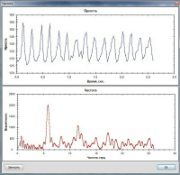
| Ciliated epithelium, video, object markers are set on the first frame. |
|
A graph depicting brightness fluctuation inside the object serves as a tool for precise marker positioning: presence of a distinguished pike means a successful measurement spot. |
|
Dependency graph: parameter vs. time. |
|
|
| |
|
|
| |
MMC MultiMeter software for microscope can work with images as well as make measurements of different parameters on videos. One of the tasks for medical microscopy consists in determination of beat frequency of respiratory cilia in ciliated epithelium. Normally, cilia beat in a coordinated manner with a specific frequency range, which results in effective clearing of mucus and debris from the airways. Ineffective movement disturbs mucociliary clearance and leads to ciliary dyskinesia. It can be the cause of sinusitis, recurrent chest infections. For diagnosis of ciliary dyskinesia with MMC MultiMeter, a video of beating cilia is recorded at the frame rate sufficient for the analysis (maximum frequency that could be measured equals half of the camera frame rate). At the first video frame, measurement markers are set in the required area (at the edge of the front of beating cilia). The software detects brightness fluctuations caused by beating cilia entering and leaving the marker, and classifies the objects according to class limits set in the classifier. Sufficient Time of video recording and measurement precision can be controlled on brightness fluctuation graphs, which are automatically generated for every marker.
The same tool can be effectively used for estimation of the frequency of any processes of technological or biological origin.
|
|
| |
|
|
| |
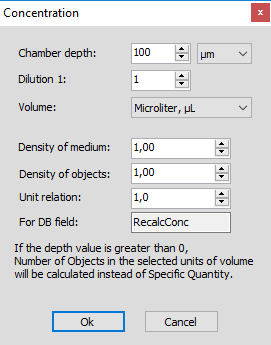
Concentration
|
|
| |
|
|
| |
|
|
The software provides special tool for estimation of concentration of objects in the specimen:
- Calculation of volume concentration and weight concentration per volume units.
- Assigning counting chamber depth.
- Considering dilution factor.
- Considering medium density and object density.
- Calculation result in chosen units of measure (grams per kilogram, grams per ton) is automatically sent to a reserved database field.
|
|
|
| |
|
|
| |
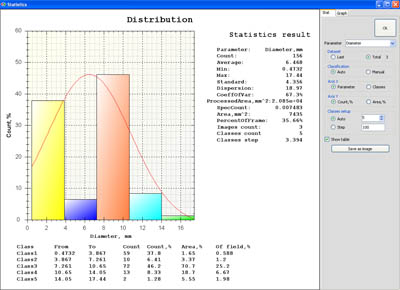
Statistics
|
|
| |
|
|
| |
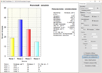
The software allows you to process a series of images and display the results with diagrams and graphs depicting dependency of different parameters from each other. Any results of statistical treatment can be sent to numerical fields of your database that are selected in statistics-database interaction settings. Images of graphs and diagrams can be used in your report. Some statistical parameters available in MMC MultiMeter:
- Object count.
- Arithmetic average value of the selected parameter.
- Minimum parameter value.
- Maximum parameter value.
- Standard deviation.
- Dispersion of parameter values.
- Variation coefficient.
- Specific quantity of objects per area unit.
- Measured area, the sum of areas of all images processed in the given series.
- Percentage of object areas vs. measured area.
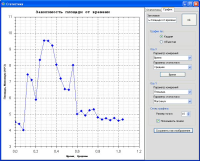
To estimate interdependency of measured parameters and assess dynamic processes on video clips, MMC MultiMeter cna build a dependency graph. If you are using parameter against time graph, the points on the graph can be connected for better visual perception.
Combining the results of microscopic analysis of a series of images, a wide range of tasks can be sold. Should you need some help in particular application, feel free to contact us with description of your task.
|
|
| |
|
|
| |
CAMERA+SOFTWARE KITS |
|
| |
-
MMC-31C12-M 3 Mp color CMOS camera is the most universal solution for any laboratory tasks in histology, cytology, geology, materials science where assessment of morphological or optical parameters
of objects is necessary. Suitable for both high resolution still images and high fps video clips recording.
- MMC-13M-M 1.3 Mp monochrome CMOS camera with 25 fps at full resolution is the best solution for analysis of moving or live objects.
- MMC-50M-M/MMC-50C-M monochrome and color 5 Mp CMOS cameras for precise details under lower magnification.
- MMC-60M-C/MMC-60C-C monochrome and color 6 Mp CMOS cameras with largest 1" sensor in our line for largest field of view and most precise details.
- MMC-14M-S/MMC-14C-S monochrome and color 1.4 Mp CCD cameras for fluorescence imaging, demanding scientific research under low light conditions.
|
|
|
|
| |
|
|
| |
|
|
|
|
| |
|
|
| |
|
|
|
|
